


Welcome to our app recommend. 20,000+ users downloaded Musculoskeletal X- Rays latest version on 9Apps for free every week! It works great even on 2G. This hot app was released on 2019-12-13. It is a very popular app on India now.
Musculoskeletal X- Rays Interpretation
chest x ray interpretation
dental x ray interpretation
x-ray interpretation
x-ray interpretation guide
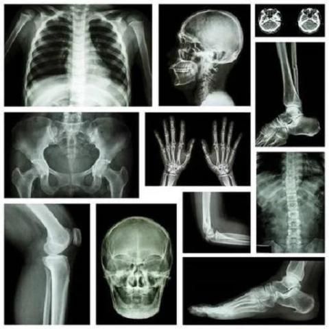
Musculoskeletal X-ray - General principles
Systematic approach
Patient and image details
Bone and joint alignment
Joint spacing
Cortical outline
Bone texture
Soft tissues
Although the system for viewing X-rays of bones and joints varies depending on the anatomy being examined, there are some broad principles which can be applied in a number of situations.A systematic approach involves checking alignment of bone structures, joint spacing, integrity of bone cortex, medullary bone texture, and for abnormalities of any visible surrounding soft tissue structures.
Patient and image details
Start by checking you are looking at the correct image. The patient's details should be checked and the date and time of the X-ray noted. The skeletal system is symmetrical, so it is particularly important to be sure you are looking at the correct side.
Bone and joint alignment
Loss of alignment may be due to a bone fracture or a joint dislocation. Both are associated with soft tissue injury that may not be directly visualised.
Joint spacing
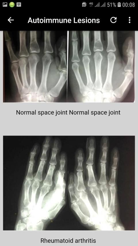
Joint spacing may be narrowed due to cartilage loss or widened due to dislocation/dissociation.
Cortical outline
Careful scrutiny of the bone cortex is required because a check that is too brief will lead to incorrect or incomplete diagnosis.In the context of trauma the clinical features of a significant injury may be masked by other injuries. Remember to be systematic, and if you spot one abnormality, do not stop until you are sure you have focused on all areas of the anatomy shown.
Bone texture
In some bones a fine matrix of fine white lines (trabeculae) is seen. Occasionally bone injury or disease will result in abnormality of this texture.
Soft tissues
Scrutinizing the soft tissues can often provide helpful information.Not uncommonly an abnormality of soft tissues is more obvious than a bone injury, or may even imply a bone injury that is not visible at all.
Confidence in assessing musculoskeletal system X-rays comes from experience and a knowledge of normal appearances. All patients are different, so being sure of the distinction between normal and abnormal is often difficult.Here are some principles that may help you to determine if a finding is normal.
2 views
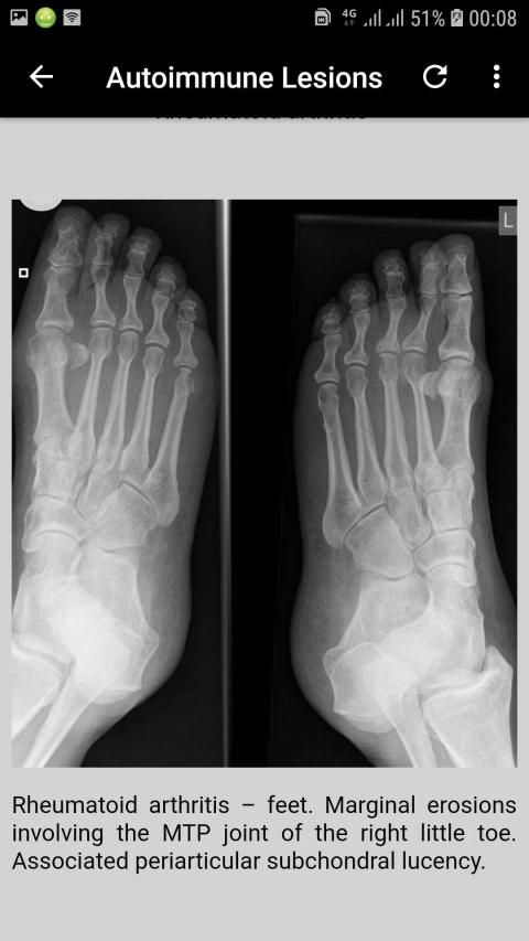
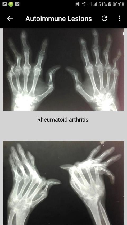
In the context of trauma at least 2 views of the body part in question are usually required. If looking for specific disease entities, for example erosions in rheumatoid arthritis, this may be less important. In some cases, such as possible scaphoid injury, more than 2 images are required.2 views are better than 1
Compare with other side
Images of the asymptomatic contralateral side to a suspected abnormality are not routinely acquired for assessment of all bones or joints.If an old image of the contralateral side is available, or if the other side is included as standard (for example hip/pelvis), then comparison between symptomatic and asymptomatic appearances can be very helpful.
Compare current with previous images
The 'old X-ray' is said to be the 'cheapest test in radiology.'
If you are uncertain of an abnormality and there is an old image available of the area in question, then ALWAYS look at it. Doing this often increases diagnostic confidence, and can show progression of pathology over time.
Keep your eye on the ball
When looking at an X-ray always keep the current clinical features at the forefront of your mind.Remember - 'Treat the patient and not the X-ray!'
Look for the unexpected
Not all disease that presents with musculoskeletal symptoms is primarily related to pathology of the bones or joints. Very often pain is referred to the symptomatic area and is explained by disease of another system.For example, shoulder pain is usually due to shoulder pathology, but always keep in mind that pain may be referred to the shoulder from the cervical spine, brachial plexus or diaphragm.
Interpretasi Sinar-X Muskuloskeletal
interpretasi rontgen dada
interpretasi x-ray gigi
interpretasi x-ray
panduan interpretasi x-ray
X-ray muskuloskeletal - Prinsip umum
Pendekatan sistematis
Rincian pasien dan gambar
Penyelarasan tulang dan sendi
Jarak sendi
Garis luar kortikal
Tekstur tulang
Jaringan lunak
Meskipun sistem untuk melihat sinar-X tulang dan sendi bervariasi tergantung pada anatomi yang diperiksa, ada beberapa prinsip luas yang dapat diterapkan dalam sejumlah situasi. Pendekatan sistematis melibatkan pengecekan keselarasan struktur tulang, jarak sendi, integritas korteks tulang, tekstur tulang meduler, dan untuk kelainan struktur jaringan lunak yang terlihat di sekitarnya.
Rincian pasien dan gambar
Mulailah dengan memeriksa Anda melihat gambar yang benar. Rincian pasien harus diperiksa dan tanggal serta waktu rontgen dicatat. Sistem kerangka simetris, jadi sangat penting untuk memastikan Anda melihat sisi yang benar.
Penyelarasan tulang dan sendi
Hilangnya keselarasan mungkin karena patah tulang atau dislokasi sendi. Keduanya berhubungan dengan cedera jaringan lunak yang mungkin tidak secara langsung divisualisasikan.
Jarak sendi
Jarak sendi dapat dipersempit karena kehilangan tulang rawan atau melebar karena dislokasi / disosiasi.
Garis luar kortikal
Pengawasan korteks tulang yang hati-hati diperlukan karena pemeriksaan yang terlalu singkat akan mengarah pada diagnosis yang tidak benar atau tidak lengkap. Dalam konteks trauma, gambaran klinis cedera yang signifikan dapat ditutupi oleh cedera lain. Ingatlah untuk sistematis, dan jika Anda menemukan satu kelainan, jangan berhenti sampai Anda yakin Anda telah fokus pada semua area anatomi yang ditunjukkan.
Tekstur tulang
Dalam beberapa tulang matriks halus garis putih halus (trabekula) terlihat. Kadang-kadang cedera atau penyakit tulang akan menyebabkan kelainan tekstur ini.
Jaringan lunak
Memperhatikan jaringan lunak seringkali dapat memberikan informasi yang bermanfaat. Tidak jarang kelainan jaringan lunak lebih jelas daripada cedera tulang, atau bahkan menyiratkan cedera tulang yang tidak terlihat sama sekali.
Keyakinan dalam menilai sistem muskuloskeletal sinar-X berasal dari pengalaman dan pengetahuan tentang penampilan normal. Semua pasien berbeda, jadi memastikan perbedaan antara normal dan abnormal seringkali sulit. Berikut adalah beberapa prinsip yang dapat membantu Anda menentukan apakah temuan itu normal.
2 kali dilihat
Dalam konteks trauma setidaknya diperlukan 2 pandangan bagian tubuh yang dipertanyakan. Jika mencari entitas penyakit tertentu, misalnya erosi pada rheumatoid arthritis, ini mungkin kurang penting. Dalam beberapa kasus, seperti kemungkinan cedera skafoid, diperlukan lebih dari 2 gambar.2 tampilan lebih baik daripada 1
Bandingkan dengan pihak lain
Gambar sisi kontralateral asimptomatik dengan dugaan kelainan tidak diperoleh secara rutin untuk penilaian semua tulang atau sendi. Jika gambar lama sisi kontralateral tersedia, atau jika sisi lain dimasukkan sebagai standar (misalnya pinggul / panggul), maka perbandingan antara penampilan simtomatik dan asimptomatik bisa sangat membantu.
Bandingkan saat ini dengan gambar sebelumnya
'Sinar X lama' dikatakan sebagai 'tes termurah dalam radiologi.'
Jika Anda tidak yakin dengan kelainan dan ada gambar lama yang tersedia di area tersebut, maka SELALU melihatnya. Melakukan hal ini sering meningkatkan kepercayaan diagnostik, dan dapat menunjukkan perkembangan patologi dari waktu ke waktu.
Perhatikan bolanya
Ketika melihat X-ray selalu menjaga fitur klinis saat ini di garis depan pikiran Anda. Ingat - 'Perlakukan pasien dan bukan sinar-X!'
Cari yang tak terduga
Tidak semua penyakit dengan gejala muskuloskeletal terutama terkait dengan patologi tulang atau sendi. Sangat sering nyeri dirujuk ke area simptomatik dan dijelaskan oleh penyakit sistem lain. Misalnya, nyeri bahu biasanya disebabkan oleh patologi bahu, tetapi selalu ingat bahwa nyeri dapat dirujuk ke bahu dari tulang belakang leher, pleksus brakialis atau diafragma.


9Apps 4.9


