

With the updating of mobile application, 20,000+ users downloaded CT Brain Basic Interpretation latest version on 9Apps for free every week! It is simply to learn how to use it. This hot app was released on 2019-06-27. You’ll want to use it on your own phones after you know more.
What is CT Scanning of the Head?
Computed tomography, more commonly known as a CT or CAT scan, is a diagnostic medical test that, like traditional x-rays, produces multiple images or pictures of the inside of the body.
The cross-sectional images generated during a CT scan can be reformatted in multiple planes, and can even generate three-dimensional images. These images can be viewed on a computer monitor, printed on film or by a 3D printer, or transferred to a CD or DVD.
CT images of internal organs, bones, soft tissue and blood vessels provide greater detail than traditional x-rays, particularly of soft tissues and blood vessels.
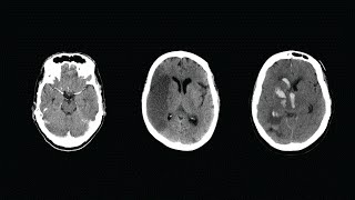
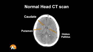
Computed tomography (CT) scanning of the head uses a series of x-rays of the head taken from many different directions; the resulting data is transformed into a series of cross sections of the brain using a computer program.
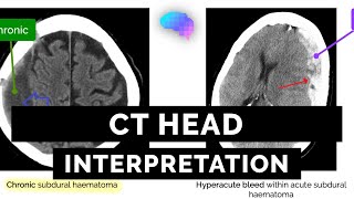
The CT head scan is one of the most common imaging studies that you can be faced with and the most frequently requested by A&E. This app will cover some of the underlying principles of CT head studies, and discuss a method for their interpretation.
Underlying principles and terminology
Computed tomography (CT) scanning involves the use of x-rays to take cross-sectional images of the body. This is possible as different tissues interact with X-rays in different ways. Some tissues will allow the passage of these X-rays without influencing it much, whilst other tissues will exert a more significant effect. The extent to which a material can be penetrated by an x-ray beam is described in terms of an attenuation coefficient which assesses how much a beam is weakened by passing through a voxel of tissue (voxel = volumetric pixel).
सिर का सीटी स्कैन क्या है?
कंप्यूटेड टोमोग्राफी, जिसे आमतौर पर सीटी या कैट स्कैन के रूप में जाना जाता है, एक नैदानिक चिकित्सा परीक्षण है जो पारंपरिक एक्स-रे की तरह, शरीर के अंदर की कई छवियों या चित्रों का उत्पादन करता है।
सीटी स्कैन के दौरान उत्पन्न क्रॉस-अनुभागीय छवियों को कई विमानों में पुन: स्वरूपित किया जा सकता है, और यहां तक कि तीन आयामी चित्र भी उत्पन्न हो सकते हैं। इन छवियों को एक कंप्यूटर मॉनीटर पर देखा जा सकता है, जो फिल्म या 3 डी प्रिंटर पर मुद्रित होता है, या सीडी या डीवीडी में स्थानांतरित किया जाता है।
आंतरिक अंगों, हड्डियों, नरम ऊतक और रक्त वाहिकाओं की सीटी छवियां पारंपरिक एक्स-रे की तुलना में अधिक विस्तार प्रदान करती हैं, विशेष रूप से नरम ऊतकों और रक्त वाहिकाओं की।
कम्प्यूटेड टोमोग्राफी (सीटी) सिर की स्कैनिंग कई अलग-अलग दिशाओं से ली गई सिर की एक्स-रे की एक श्रृंखला का उपयोग करती है; परिणामी डेटा को कंप्यूटर प्रोग्राम का उपयोग करके मस्तिष्क के क्रॉस सेक्शन की एक श्रृंखला में बदल दिया जाता है।
सीटी हेड स्कैन सबसे आम इमेजिंग अध्ययनों में से एक है जिसका आप सामना कर सकते हैं और ए एंड ई द्वारा सबसे अधिक बार अनुरोध किया जा सकता है। यह ऐप सीटी हेड अध्ययन के कुछ अंतर्निहित सिद्धांतों को कवर करेगा, और उनकी व्याख्या के लिए एक विधि पर चर्चा करेगा।
अंडरस्टैंडिंग सिद्धांतों और शब्दावली
कंप्यूटेड टोमोग्राफी (सीटी) स्कैनिंग में शरीर की क्रॉस-सेक्शनल इमेज लेने के लिए एक्स-रे का उपयोग शामिल है। यह संभव है क्योंकि विभिन्न ऊतक अलग-अलग तरीकों से एक्स-रे के साथ बातचीत करते हैं। कुछ ऊतक इन एक्स-रे के पारित होने की अनुमति देगा बिना इसे प्रभावित किए, जबकि अन्य ऊतक अधिक महत्वपूर्ण प्रभाव डालेंगे। एक एक्स-रे बीम द्वारा एक सामग्री को किस हद तक प्रवेश किया जा सकता है इसका वर्णन एक क्षीणन गुणांक के संदर्भ में किया जाता है जो यह आकलन करता है कि ऊतक के एक voxel (voxel = volumetric पिक्सेल) से गुजरने से कितना बीम कमजोर होता है।
New release.
9Apps 4.9